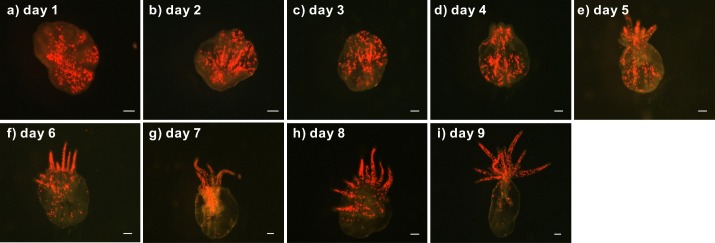
Figure 2. Development of a representative lacerate collected from a clade C Symbiodinium-infected anemone.
First, the lacerate was transferred to a new dish immediately following laceration from the pedal disk of a clade C Symbiodinium-infected anemone. The development of the lacerate and spread of Symbiodinium was recorded daily using a fluorescent stereomicroscope (A–I). The red spots in the images indicate chlorophyll autofluorescence of the Symbiodinium cells. Scale bars: 100 µm.

