
Figure 2.
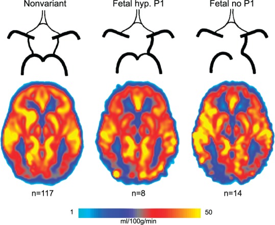
Mean pCASL-CBF maps for the left-sided cases of the three most prevalent posterior circle of Willis variants. A reduction in pCASL-CBF signal can be seen on the posterior side contralateral to the fetal-type posterior cerebral artery. Fetal hyp. P1: fetal variant with hypoplastic ipsilateral P1 segment; Fetal no P1: fetal variant lacking the ipsilateral P1 segment.