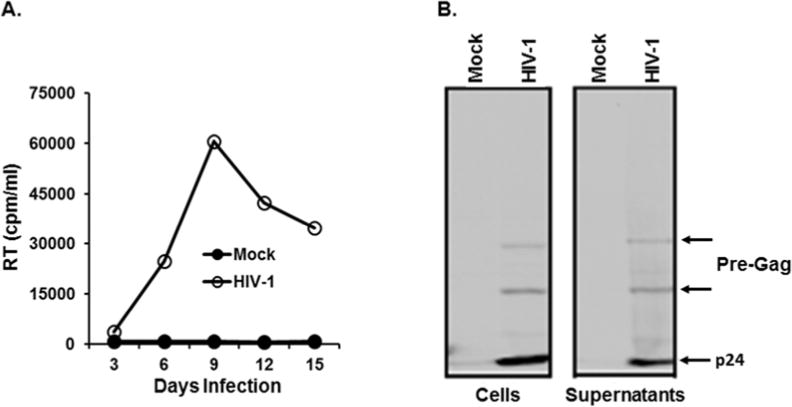
Figure 1.

Replication of HIV-1 in Jurkat and WB analysis of viral proteins. (A) HIV-1 corresponding to 10,000 cpm/ml was infected into Jurkat cells (1×10⁶ cells), and replication kinetics of HIV-1 was determined by measuring RT activity in the culture supernatants every 3 days. (B) HIV-1 was infected into Jurkat cells, and cell (left) and virus (right) lysates were prepared at peak virus replication. 1/5 and 1/2 of total lysates of cells and virus (supernatants), respectively, were employed for the analysis. Band intensity of p24 (arrow) in the supernatant was approximately 1/3 of that in the cells.
