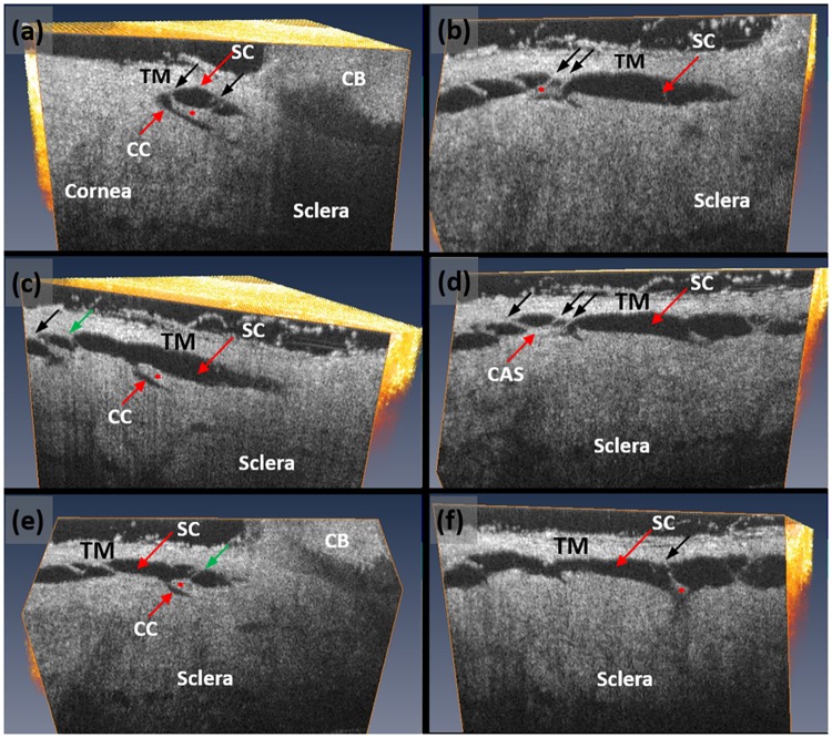
Fig 4. High-resolution volumetric SD-OCT dataset permits detailed examination of the trabecular meshwork (TM), Schlemm’s canal (SC) (red arrow) and the collector channels (CC) (white arrow).
Images shown are obtained from dissecting the scanned tissue volume at a plane that gives optimal identification of collagen flaps to show the hinged flap or leaflet-like organization at the collector channel entrance. (a-f) Images are each oriented to provide an optimal view of collector channel relationships to SC, and the associated hinged collagen flaps (asterisks) that separate the collector channel from SC.). Each CC has a relatively long flap at its entrance creating the appearance of a hinged configuration. The hinged flaps or leaflets are each attached to the TM by means of thin cylindrical attachment structures (CAS) (black arrows) spanning SC. Some sections through the CAS revealed the presence of a lumen (green arrows).

