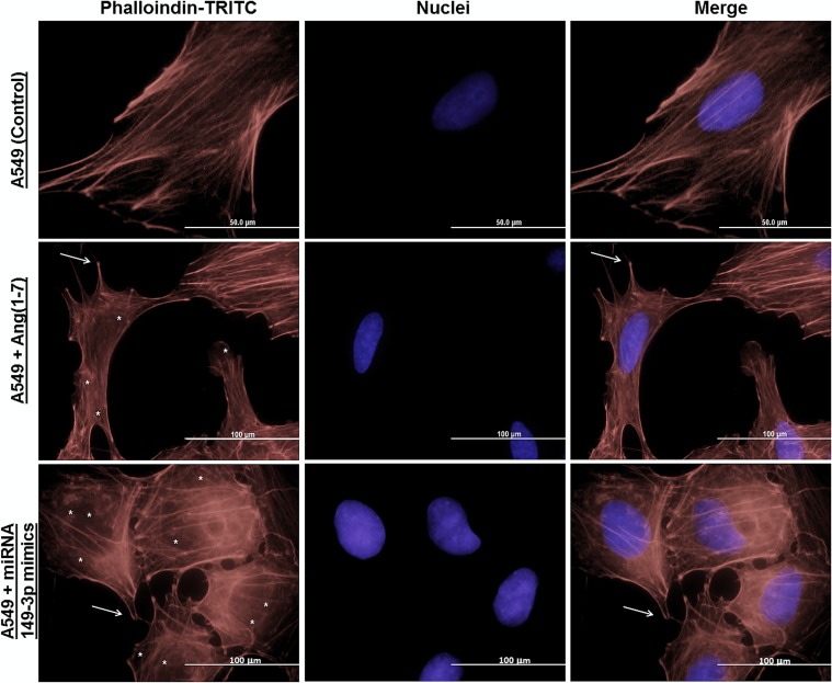
Fig 3. Immunocytochemistry of actin filaments of A549 cells.
Different groups of A549 cells were grown on coverslips and then stained with phalloidin-TRITC and DAPI (nuclei) to investigate morphologic changes in cellular groups. Arrows (→) indicate filopodia and asterisks (*) indicate actin filaments disassemble in the images. Scale bar are assigned.

