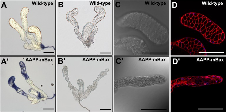
Fig 3. Salivary glands of an AAPP-mBax mosquito.
(A, A’) Salivary glands of 3-day-old adult female wild-type mosquitoes and AAPP-mBax mosquitoes stained with trypan blue. (B) A normal salivary gland from a 7-day-old adult female wild-type mosquito. (B’) An abnormal salivary gland with aberrant distal-lateral lobes from a 7-day-old adult female AAPP-mBax mosquito (line 1). (C, C’) Differential interference contrast images of the salivary glands of a 7-day-old adult female wild-type mosquito and AAPP-mBax mosquito (line 1). (D, D’) Fluorescence images of the salivary glands of a 7-day-old adult female wild-type mosquito and AAPP-mBax mosquito stained with CellMask (Red) as a marker for cell membranes and Hoechst 33342 (Blue) as a marker for DNA. Scale bars = 100 μm.

