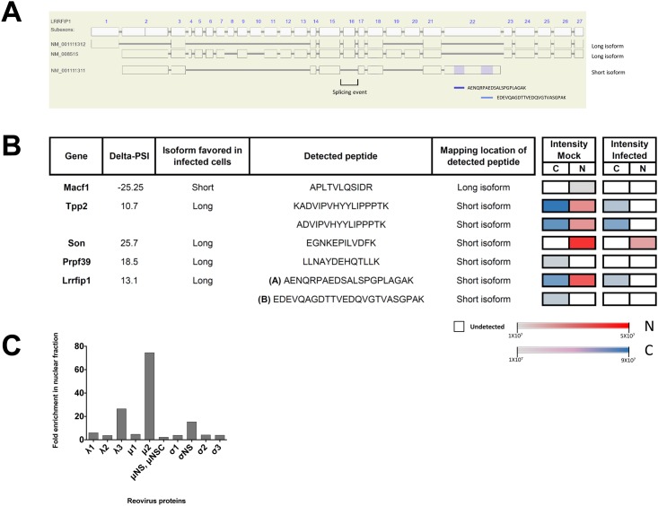
Fig 5. Proteomic analysis of uninfected and reovirus-infected cells.
(A) LRRFIP1 coding exon structure and localization of peptides detected for this gene. Two long isoform transcripts (NM_001111312; NM_008515) and one short isoform transcript (NM_001111311) are also represented. Other short (BC144955; AK044174) and long (BC145642) isoforms analyzed in the RNA-Seq process were omitted. (B) Transcript-specific peptides detected for the long/short transcript. The intensities of peptide detection for both uninfected (mock) and infected cells in the cytoplasm/nucleus fractions are displayed as color gradation (Cytoplasm: grey = weakly detected, blue = strongly detected, white = undetected; Nucleus: grey: lightly detected, red = strongly detected; white = undetected). For Lrrfip1, two peptides (A and B) are presented. (C) Fold enrichment of reovirus proteins in nuclear fraction over the cytoplasmic one. Proteins μ2 (74.5x), λ3 (26.5x) and σNS (15.4x) were found to be above the background level of other viral proteins.

