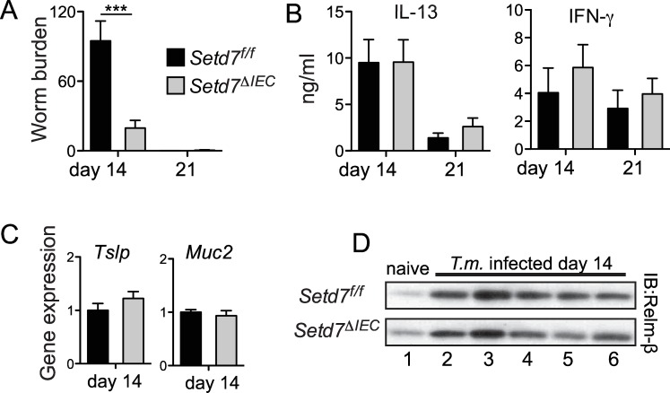
Fig 2. Deletion of Setd7 specifically in IECs renders mice resistant to T. muris.
(A) Setd7 f/f (black bars) and Setd7 ΔIEC (grey bars) mice were infected with 200 T. muris eggs and worm burdens were determined at day 14 or day 21 post infection (n≥9 for day 14, n = 6 for day 21, *** P<0.001). (B) Mesenteric lymph node cells from infected mice were re-stimulated for 72 h. IL-13 and IFN-γ concentrations in supernatants was determined by ELISA. (n≥4). (C) Expression of Tslp and Muc2 in proximal colon at day 14 post infection with T. muris was assessed by qPCR. Expression is relative to infected control (Setd7 f/f) mice. (n≥8). (D) Protein levels of RELMβ that was secreted into the gut lumen of naïve and T. muris infected mice evaluated by Western blot.

