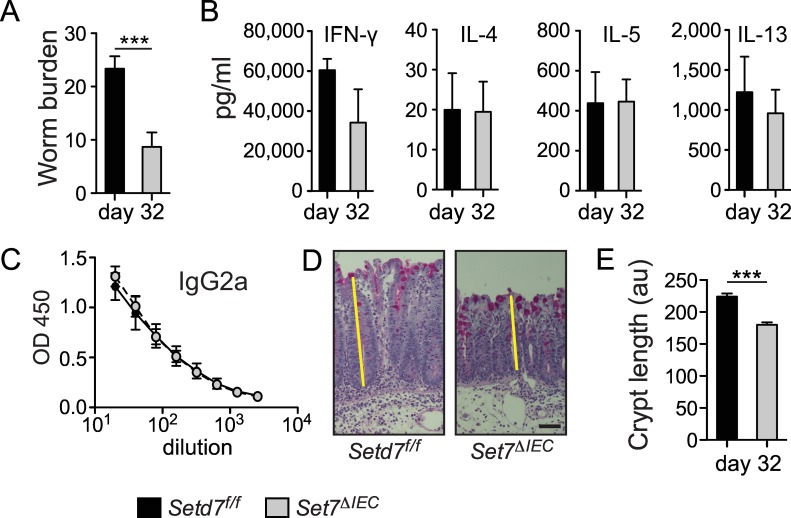
Fig 4. Setd7 ΔIEC mice resistant to low dose T. muris infection.
(A) Setd7 f/f and Setd7 ΔIEC mice were infected with ~35 T. muris eggs (low dose) and worm burdens were determined at day 32 post infection (n = 9, pooled from 2 independent experiments, *** P<0.001). (B) Cytokine production of mesentyric lymph node cells that were re-stimulated for 72 h was determined by ELISA. (n≥4). (C) Serially diluted serum of infected mice was analyzed by ELISA to measure T. muris-specific IgG2a. (n≥4). (D) Periodic acid-Schiff stained caecal sections show a robust depletion of goblet cells in both Setd7 f/f and Setd7 ΔIEC mice at day 32 post infection. Yellow lines indicate crypt length. Original magnification is 100X. Bar = 50 μm (E) Crypt length (au = arbitrary units) in caecums of mice at day 32 post infection. (n≥60 of 9 mice in each group, *** P<0.001). Setd7 f/f black bars, Setd7 ΔIEC grey bars.

