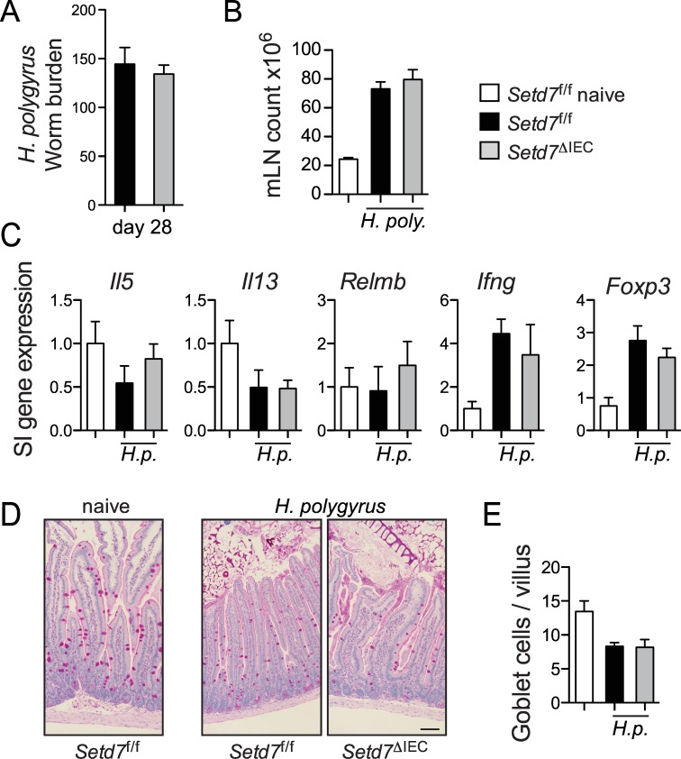
Fig 5. Setd7 ΔIEC mice do not have increased resistance against infection with H. p. bakeri.
(A) Setd7 f/f and Setd7 ΔIEC mice were infected with ~ 200 H. p. bakeri eggs and were killed day 28 post infection. Worm burdens were determined microscopically from the small intestine. (B) Mesenteric lymph node (mLN) cell counts from naïve and H. p. bakeri infected mice (day 28 post infection). (C) Gene expression of indicated genes in small intestine (SI) that was adjacent to infection site at day 28 post infection with H. p. bakeri. Expression is relative to naïve mice (white bars). (D) Periodic acid-Schiff (PAS) staining of small intestinal tissue sections of indicated mice at day 28 post infection. Bar = 100 μm. (E) PAS+ cells per villus were counted from images as shown in (D). (A-E) n = 3 (naives), n = 8 (infected) from 2 independent experiments. Setd7 f/f black bars, Setd7 ΔIEC grey bars.

