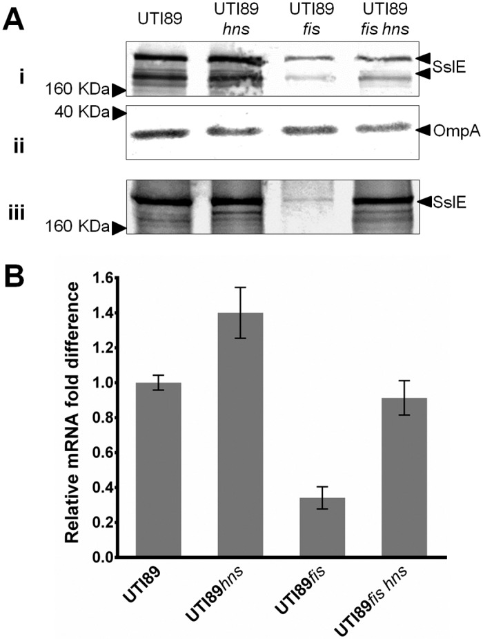
Fig 6. Effect of fis and hns double deletion on sslE expression in UTI89 determined via western blot and qRT-PCR analysis.

(A) Western blot analysis of SslE using (i) whole-cell lysates and (iii) supernatant fractions prepared from UTI89, UTI89hns, UTI89fis and UTI89fis hns. (ii) Western blot loading control for whole cell lysate samples using an OmpA antibody. The overall level of SslE was reduced in UTI89fis hns compared to wild-type UTI89. (B) Relative fold-difference of sslE transcript levels of UTI89, UTI89hns, UTI89fis and UTI89fis hns. All mRNA levels were calculated relative to the level of UTI89 sslE mRNA. The relative sslE mRNA levels were consistent with the data observed from the western blot analysis. The data was obtained from three independent experiments; error bars indicate standard deviation.
