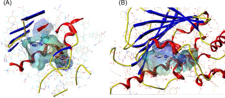
Fig. 3.
The comparison of ATP binding site of (A) topo II and (B) Hsp90. The channel was created using MOLCAD implemented in Sybyl, colored by electrostatic potential. The color ramp ranges from red (most positive) to purple (most negative). AMPPNP and ADP bound to topo II and Hsp90, respectively are represented in sticks colored by atom type (gray: carbon; red: oxygen; blue: nitrogen; orange: phosphorus). The proteins are represented in ribbon (blue: β-strand; red: α-helix).

