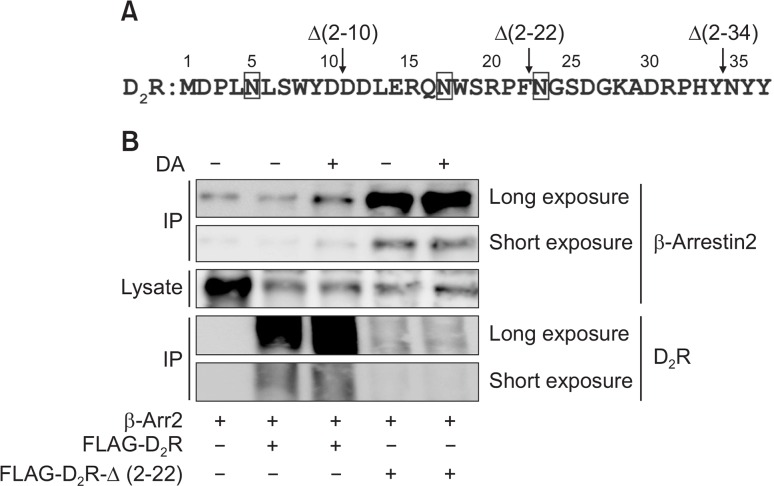
Fig. 3.
Role of the N-terminal region of D2R in the interaction with β-arrestin2. (A) Alignment of the amino acid sequences within the N-terminal regions of D2R. The numbers represent the position of the amino acid residues starting from Met1. Vertical arrows show the positions at which deletions were made. Squares represent consensus N-linked glycosylation sites. (B) Effects of shortening the N-terminal region on the interaction between D2R and β-arrestin2. HEK-293 cells were transfected with 0.8 μg of Flag-tagged D2R and 8 μg Flag-D2R-Δ(2–22) in pCMV5 per 100 mm culture dishes. Cells were treated with 10 μM DA for 5 min. Cell lysates were immunoprecipitated with Flag beads, and analyzed by an immunoblot assay with antibodies against β-arrestin2 or Flag. Cell lysates were immunoblotted with antibodies against β-arrestin2. Data represent results from three independent experiments with similar outcomes.

