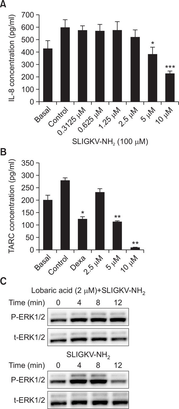
Fig. 2.
Lobaric acid decreased PAR2-induced inflammatory response in keratinocytes. After pretreatment with lobaric acid, 100 μM SLIGKV-NH2 was added to HaCaT keratinocytes for 24 hours and immunoassays were performed (A). INF-γ and TNF-α were added to HaCaT keratinocytes followed by lobaric acid or dexamethasone. Supernatants were used in immunoassays (B). ERK phosphorylation in NHEKs induced by 100 μM SLIGKV-NH2 was blocked by lobaric acid. NHEKs were stimulated for indicated times with 100 μM SLIGKV-NH2 and stimulation was blocked by pretreatment with 2 μM lobaric acid (C). Whole cell lysates were used to determine ERK phosphorylation (p-ERK) and total-ERK (t-ERK) by Western blots. Data are mean ± SEM, n=2. *p<0.05, **p<0.01, ***p<0.005, compared with controls.

