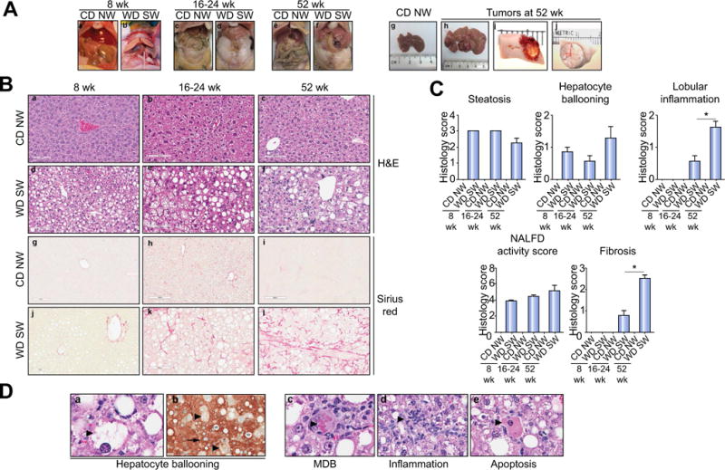
Fig. 2. DIAMOND mice sequentially develop a fatty liver, steatohepatitis, advanced fibrosis and liver tumors.

(A) Gross liver from B6/129 mice fed a chow diet (CD NW) or high fructose/glucose, high fat Western Diet (WD SW) for 8 (a, b), 16–24 (c, d) and 52 weeks (e, f). In mice fed a high fat Western Diet for 52 weeks, multiple foci of tumors were observed at the time of necropsy (h–j) as compared to CD NW mice (g); areas of hemorrhage were seen in larger tumors (i). (B) Microscopic views of livers from CD NW or WD SW mice at 8 (a, d, g, j), 16–24 (b, e, h, k) or 52 weeks (c, f, i, l) of diet. Representative liver sections stained with hematoxylin-eosin (H&E) (a–f) or Picrosirius Red (g–l) are shown. Original magnification, ×20. (C) Histology score for steatosis, hepatocyte ballooning, lobular inflammation, NAFLD Activity Score and fibrosis were quantified. Data are expressed as the mean ± SEM for 6–10 mice per group; *p <0.05. (D) Representative images of hematoxylin-eosin (H&E) (a, c–e) and CK-18 (b) staining of liver tissue from mice fed a WD SW for 52 weeks depicting the individual component of steatohepatitis (as indicated by large arrow): (a) hepatocyte ballooning, (c) Mallory-Denk bodies (MDB), (d) lobular inflammation and (e) apoptotic bodies. (b) A marked CK-18 staining is present in the cytoplasm of normal hepatocytes (as indicated by small arrow), whereas in ballooned hepatocyte (as indicated by large arrow), a reduction (almost a complete loss) in CK-18 staining in their cytoplasm is noted. Lipid vacuoles are indicated by a star. Original magnification, ×40.
