
Fig. 3. Main histological features of DIAMOND mice are comparable to human NASH.
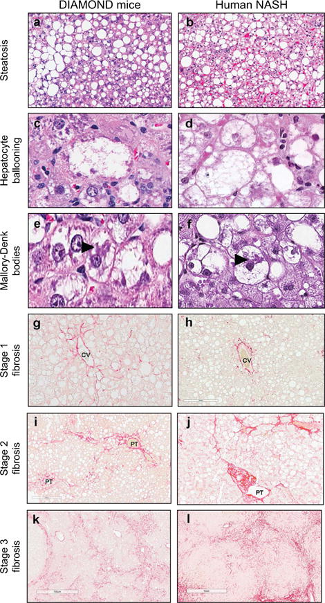
Representative images of liver histology from DIAMOND mice (a,c,e,g,i,k) or human NASH (b, d, f, h, j, l) depicting steatosis (a, b; H&E; original magnification, ×5), Hepatocyte ballooning (c, d; H&E; original magnification, ×40), Mallory-Denk bodies (as indicated by large arrow) (e, f; H&E; original magnification, ×40), Stage 1 fibrosis (g, h; Sirius Red; original magnification, ×20), Stage 2 fibrosis (i, j; Sirius Red; original magnification, ×20), Stage 3 fibrosis (k, l; Sirius Red; original magnification, ×10). CV, central vein; PT, portal tract.