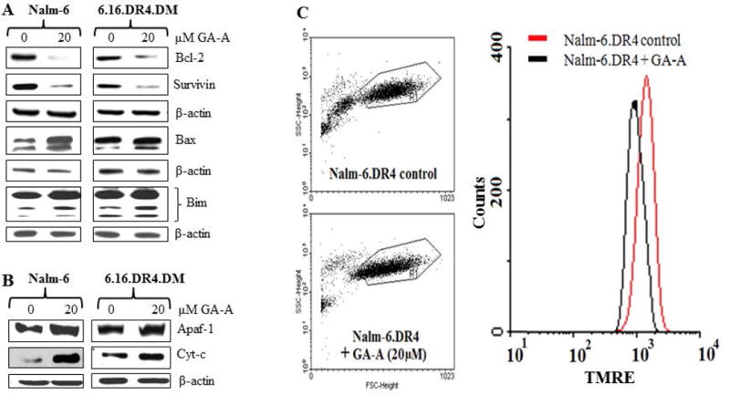
Fig. 3.

GA-A treatment alters apoptosis regulatory molecules in B-lymphoma cells. (A) Western blot analysis of whole cell lysates showing suppression of anti-apoptotic molecules BCL-2 and survivin, and an up-regulation of pro-apoptotic BIM, BAX proteins in NALM-6.DR4 and 6.16.DR4.DM cells treated with GA-A (0 and 20μM) for 24 h. (B) Western blot analysis showing an up-regulation of APAF-1 and cytochrome c proteins in enriched cytoplasmic fractions of NALM-6.DR4 and 6.16.DR4.DM cells. Cells were treated with vehicle alone or GA-A for 24h, and were subjected to Western blotting for BCL-2, Survivin, BAX, BIM, APAF-1 and cytochrome c proteins. β-actin was used as a loading control. (C) Flow cytometry analysis of TMRE stained NALM-6.DR4 cells after 20μM of GA-A treatment for 24h at 37°C. Left panels show dot plots of forward and side scatter (20,000 events/sample) obtained from control and GA-A-treated cells. Right panel shows histograms extracted from dot plots of TMRE staining of control (red line) and GA-A treated (black line) cells. Data are representative of at least three independent experiments.
