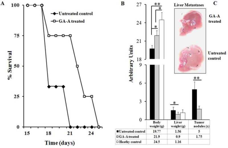
Fig. 5.

GA-A treatment reduces metastatic growth of lymphoma in vivo. Syngeneic male mice were injected with EL4 cell line as described in Materials and Methods. (A) Figure showing survival study of GA-A treated and untreated control mice. Eight mice were used in each treatment group. Results are expressed as a percentage of the initial group surviving at each time point (average survival for GA-A treated and untreated control was 22.25 days vs. 18 days, respectively; n = 8, p = 0.0003). (B) Average weights of whole body and liver, as well as the average number of tumor nodules were counted on the surface of liver samples and expressed as arbitrary units. Quantitative data were shown in the table below. (C) A representative photograph of liver samples collected from GA-A and vehicle treated animals, showing the abundance of tumor nodules in untreated controls (arrows). Significant differences were calculated by student’s t-test; **p<0.01.
