
Figure 1.
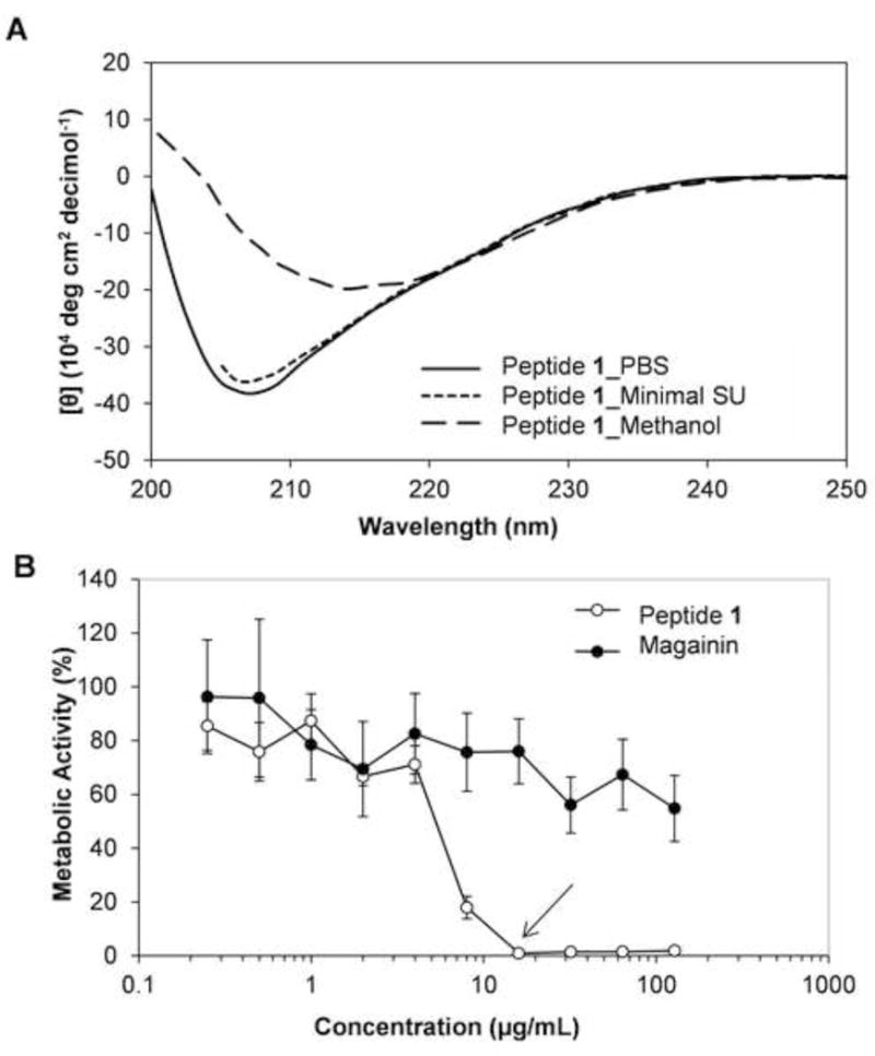
(A) Circular dichroism spectra of β-peptide 1 in PBS (solid line), in minimal SU medium (dotted line), and in methanol (dashed line). (B) Planktonic antifungal activity of β-peptide 1 (solid circles) and antimicrobial α-peptide magainin-2 (open circles) in SU media. C. albicans cells (103 cells/mL) were incubated in the presence of 2-fold dilutions of peptide for 48 hours and XTT was used to assess metabolic activity. Data points are averages of at least two independent experiments with triplicates in each and error bars denote standard deviation. Arrow indicates MIC of the peptide in SU medium.