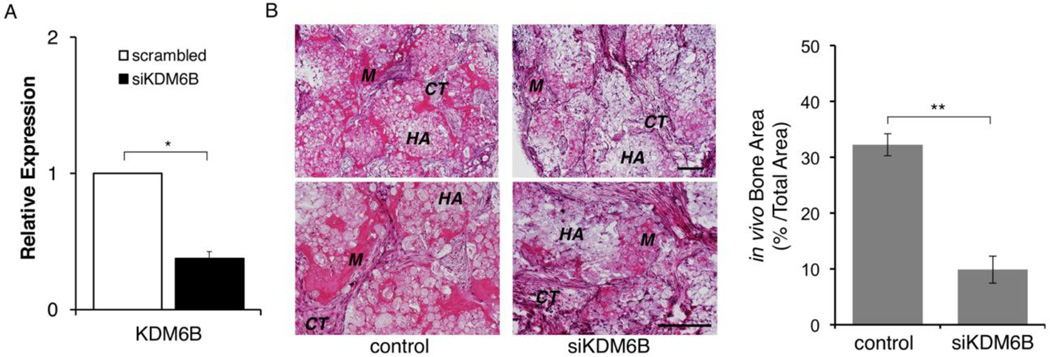
Figure 5. Knockdown of KDM6B resulted in a reduced mineralization potential in vivo.
A. DPSCs were transiently transfected with a scrambled siRNA or siKDM6B construct (Thermo Scientific, Waltham, MA). Quantitative RT-PCR analysis showed >2-fold reduction in KDM6B in siKDM6B cells. Error bar shows the standard error margin (SEM). Statistical significance was determined by Student t-test (*: p<0.05). B. DPSCs transfected with control (scrambled siRNA) or siKDM6B cells were implanted into mice as described in Materials and Methods. Formation of mineralized tissue (M) and connective tissue (CT) around HA/TCP (HA) are indicated in H&E staining section. Quantitative measurement showed that siKDM6B resulted in about 66% reduction in bone area compared to the control. Statistical significance was determined by Student t-test (**: p<0.05). Error bar shows the standard error margin (SEM).

