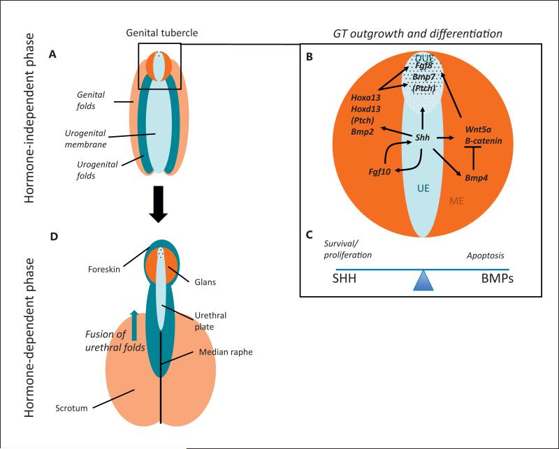
Fig. 2.
The molecular pathways underlying the development of the male external genitalia. A–C The hormone-independent phase where the urogenital sinus is indifferent. This phase occurs following the division of the cloacal membrane into the urogenital sinus and anal membrane. A At this stage the tissue is made up of the urethral folds (dark blue), the urethral epithelium (light blue), the labioscrotal swellings (light orange), and the mesenchyme of the GT (dark orange). B The GT (enlarged in box) consists of a urethral plate epithelium (UE), including the distal urethral epithelium (DUE), surrounded by a bilateral mesenchyme (ME). Numerous signalling molecules contribute to GT outgrowth and differentiation during this first phase. A central cue is SHH, which is expressed in the UE, and activates its pathway via its receptor Patched (PTCH). In the bilateral mesenchyme the HH pathway activates the expression of Hoxa13 and Hoxd13, Wnt5a and its pathway (including β-catenin). SHH signalling also activates Fgf8 and Bmp7 in the DUE. SHH has a positive feedback loop with FGF10 in the mesenchyme. SHH signalling is required for both GT outgrowth and cell survival. SHH targets Fgf8 and Wnt5a are also necessary for outgrowth. Opposing this, BMP4 (and other BMPs) repress outgrowth, perhaps via their repression of Wnt5a and Fgf8. C BMP4 also represses proliferation while activating cell apoptosis, a balance that is counteracted by SHH signaling. D The later hormone-dependent stage of development after masculinisation has occurred. During masculinisation, testosterone secreted by the testis causes the GT to elongate and urogenital membrane to transform into urethral groove. Following this, all components of the penis start to close ventrally, giving rise to the median raphe. A balance between testosterone and oestrogen signalling regulates these processes. The colour of each tissue represents its origin in the previous stages.

