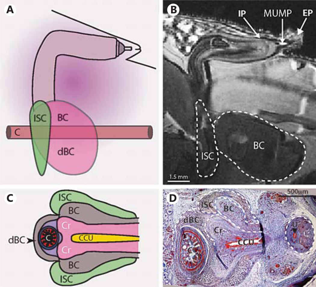
Fig. 6.
A Diagram showing the adult mouse penis from a sagittal view as it sits in the abdomen in the flaccid state. This view is also seen in B as an MRI still frame from the whole-mount penis in situ (P43) scans. Dashed lines outline the BC and ischiocavernosus muscle. The location of the IP, EP and MUMP are indicated. C Diagram of the transverse view of the terminal penis and its muscle attachments. This view is seen also in D Mallory’s trichrome staining of a P0 transverse section. Dashed lines outline the various muscles and terminating penile body, while the dashed line circle on the right outlines proximal penile G. C = Colon; Cr = crura, the terminus of the penile body; dBC = dorsal bulbocavernosus muscle; ISC = ischiocavernosus muscle; U = urethra (black arrowhead). For further abbreviations, see figures 1 and 2.

