
Figure 3.
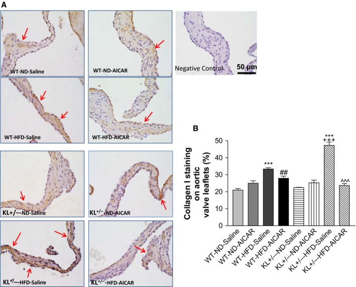
Klotho deficiency promoted upregulation of collagen I expression in aortic valves via downregulation of AMPKα in mice fed with HFD. (A) IHC staining of type I collagen (also known as collagen I) in the aortic valves of WT and Klotho‐deficient (KL +/−) mice fed with a HFD for 13 weeks followed by treatment with AICAR for an additional 2 weeks. The collagen I deposition (brown) is significantly increased in the leaflets in KL +/− mice fed with a HFD. Red arrows indicate collagen I staining (brown color) on the surface of the leaflets. (B) Quantification of collagen I level in leaflets (N = 4–6). Data = means ± SEM. ***P < 0.001 vs. WT‐ND‐Saline; ## P < 0.01 vs. WT‐HFD‐Saline; +++ P < 0.001 vs. KL +/−‐ND‐Saline, ^^^P < 0.001 vs. KL +/−‐HFD‐Saline.