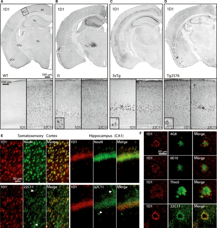
Figure 2.

Immunohistochemistry reveals mouse line‐specific hAPP expression. (A) In brains of wild‐type (WT) mice, the hAPP‐specific antibody 1D1 does not label any structures, whereas 22C11 labels numerous neurons. (B) In the brain of I5 mice, numerous neocortical neurons are labelled by the hAPP‐specific antibody 1D1. The transgene is specifically expressed by many neocortical neurons, hippocampal CA2/3 and dentate gyrus neurons as well as neurons in thalamus. (C) In 3xTg mice, the hAPP expression in neocortex is restricted to layer V pyramidal neurons and in hippocampus to CA1 and to a lesser extent CA2/3 neurons. (D) In Tg2576 mice, hAPP is expressed by neurons across all neocortical layers and in hippocampal CA1 to 3 regions. Additionally, 1D1 labels numerous plaques in the piriform cortex. (E) Top: In Tg2576 brain, double immunofluorescent labelling of hAPP by 1D1 and neurons by NeuN reveals the neuron‐specific expression of hAPP in somatosensory cortex (left) and hippocampus (right). Bottom: In Tg2576 mice, double immunofluorescent labelling of hAPP by 1D1 and mouse/human APP/APLP‐2 by 22C11 demonstrates the labelling of non‐neuronal structures by 22C11 (arrowheads) but not by 1D1 in somatosensory cortex (left) and hippocampus (right). The scale bars in the images apply to all corresponding microphotographs. The scale bar in the inset in (B) represents 20 μm. (F) Double labelling of hAPP by 1D1 (red) in combination with the Aβ‐specific antibodies 4G8 and 6E10, with thioflavin S (ThioS) and with the N‐terminal antibody 22C11 (all in green). Note the labelling of the plaque core by 6E10, 4G8 and ThioS and the labelling of dystrophic neurites in the plaque periphery by 1D1. The 1D1 antibody generates an identical labelling as 22C11. The scale bar represents 50 μm and applies to all images. sCx somatosensory cortex; RS retrosplenial cortex; hc hippocampus; CPu caudate putamen; Th thalamus, pCx piriform cortex
