Figure 3.

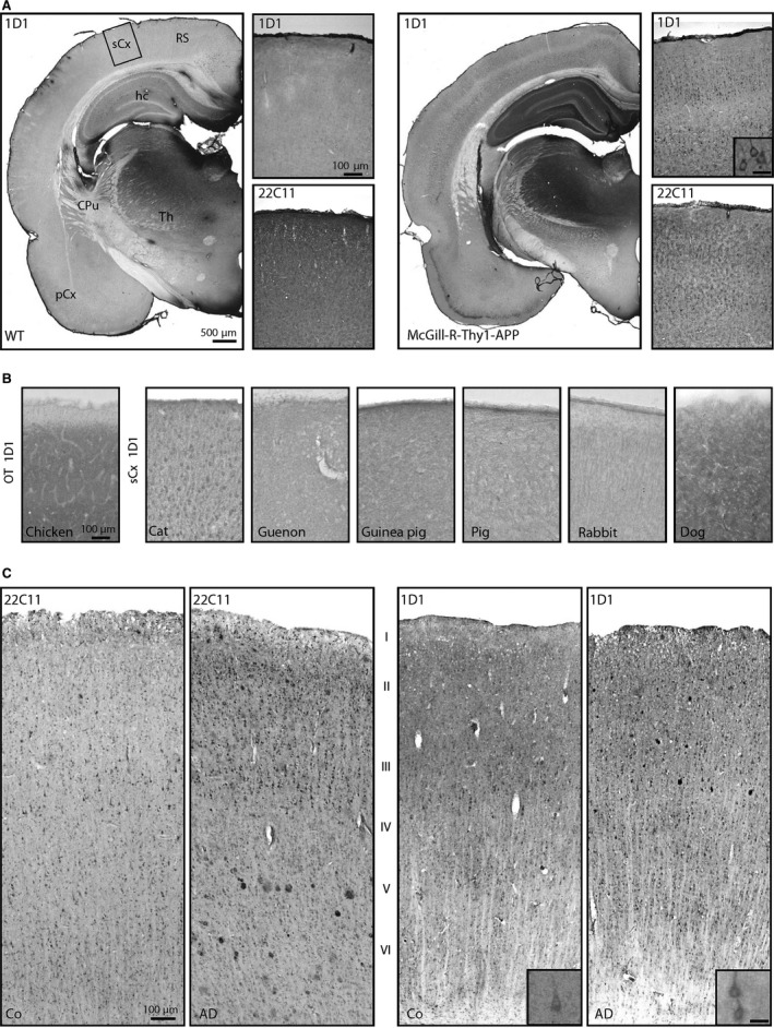
Immunohistochemistry for hAPP in hAPP‐transgenic rats, nontransgenic animal species and human control and AD brain. (A) In the brain of McGill‐R‐Thy1‐APP rats (right), numerous neocortical neurons are labelled by the hAPP‐specific antibody 1D1. Diffuse hAPP immunoreactivity is also present in hippocampus. This labelling was not detected in brains of wild‐type (WT) littermates (left). (B) In somatosensory cortex of different nontransgenic animal species, 1D1 only generated specific signals in cat, but not in chicken, guenon, guinea pig, pig, rabbit and dog. (C) In human neocortical brain tissue from control (Co) and AD subjects, 22C11 (left) and 1D1 (right) generated similar staining patterns for neurons. Both antibodies also labelled Aβ plaques. The scale bars in the images apply to all corresponding microphotographs. The scale bar in the inset in (A) represents 20 μm and also applies to the insets in (C). sCx somatosensory cortex; OT optic tectum; RS retrosplenial cortex; hc hippocampus; CPu caudate putamen; Th thalamus, pCx piriform cortex.
