
FIGURE 1.
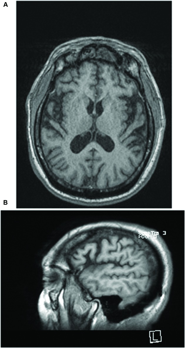
(A) T1 axial and (B) sagittal images of the brain obtained 9 months earlier demonstrate mild global volume loss for age with widening of the Sylvian fissure bilaterally. Neither evidence of past acute TBI nor white matter abnormalities were noted on fluid-attenuated inversion recovery (FLAIR) imaging.