
Figure 3.
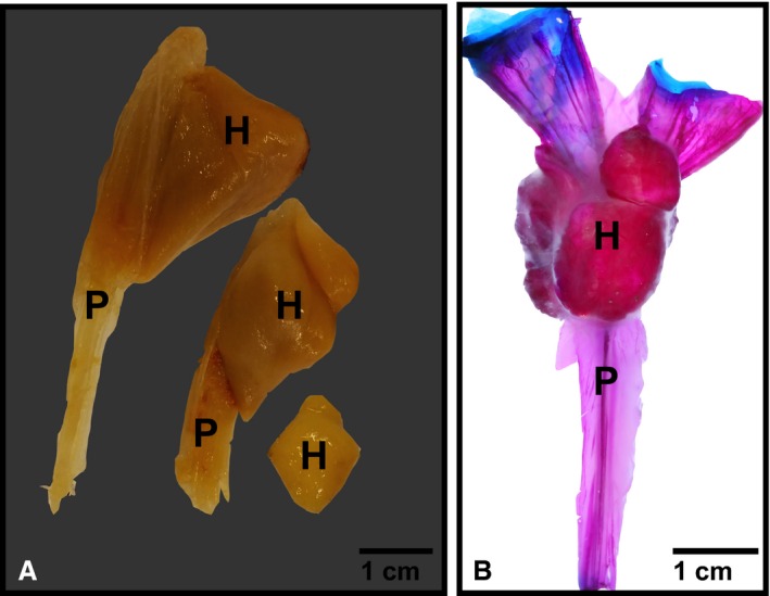
Hyperostosis in oarfish. (A) Hyperostosis along individual dorsal pterygiophores in a museum specimen (compliments of Scripps Institution of Oceanography Marine Vertebrate Collection). Specimens were already excised from the oarfish. Anterior view. (B) Cleared and stained dorsal pterygiophore from oarfish 1 (Oct 2013 specimen), lateral view. Red is mineralized tissue stained with alizarin red, blue is cartilage stained with alcian blue. Note the ascending pterygiophore (P) is poorly mineralized acellular bone compared with the cellular hyperostotic portion (H) at the distal tip. Blue stain is articular cartilage.