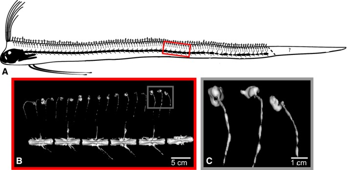
Figure 5.

Repeating, hyperossified, distal pterygiophores in adult oarfish. (A) Schematic of the scan of oarfish 1. The ‘?’ indicates the area of the tail distal to the point of autotomy. All adult scavenged fish examined were missing part of their tail. Fish 1 (this scan) shows evidence of healing on the tail suggesting the loss of the tail was not related to the stranding. (B) CT rendering of the pterygiophore hyperostoses. Some pterygiophores are bent as an artifact of freezing. (C) CT rendering of pterygiophore hyperostoses near the anal vent. The degree of hyperostosis along the pterygiophores is similar to the anterior (rostral) pterygiophores.
