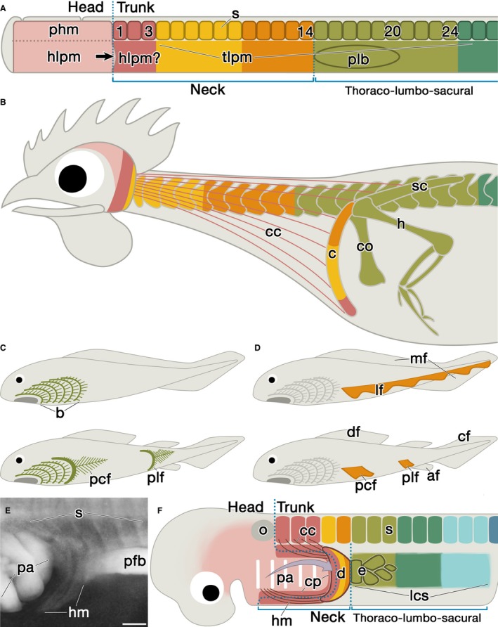
Figure 4.

Development and evolution of the neck and the pectoral appendage. (A,B) Cartoons show chickens in early embryonic (A) and adult (B) stages. Colors indicate the axial position in the early stage. Whereas paraxial head mesoderm and somites form the axial skeletons (Noden, 1984; Huang et al. 2000), LPM develops into the appendicular skeletons. The embryonic body can be divided into head and trunk regions, and the latter further into the limb‐incompetent neck region and the limb‐competent thoraco‐lumbo‐sacral region. The pectoral limb bud (plb) develops at the rostral margin of the thoraco‐lumbo‐sacral region. Note that the embryonic neck corresponds to the presumptive clavicle region in this early stage (A). The rostral part of the neck LPM could be the head mesoderm. (C,D) Classic theories on the evolution of paired appendages. The gill‐arch theory (C; Gegenbaur, 1876, 1878) posits that the appendage is made up of the transformed posterior branchial arches. The pelvic fins (plf) are assumed to have migrated caudally. The lateral fin‐fold theory (D; Thacher, 1877; Mivart, 1879; Balfour, 1881) posits that the ancestral animal possessed a paired lateral fin‐fold (lf) along the length of the trunk, and the structure divided rostrocaudally to form both pectoral (pcf) and pelvic appendages. (E) The circumpharyngeal ridge in the Scyliorhinus torazame embryo (B 27). Note that the hypobranchial muscle anlagen (hm) grow ventrorostrally between the pharyngeal arches (pa) and the pectoral fin bud (pfb). Rostral is to the left. (F) Schematic drawing showing the embryonic architecture of the neck region in late embryonic stage. The head LPM expands caudally to form the pharyngeal arches (arrow in A; Kuratani, 1997). As a result, the neck region contains the presumptive clavicle domain, the caudal pharyngeal arches, and the horseshoe‐shaped circumpharyngeal ridge (cp; reviewed by Kuratani, 1997) as their border. In the CP ridge, the caudal margin of the cephalic neural crest cells and LPM adjacent to the most rostral somites overlap. From the pharyngeal arches, the parathyroid glands develop in tetrapods and the internal gill buds in fishes (Okabe & Graham, 2004). In the CP ridge, the cucullaris and hypobranchial muscles develop (viz. circumpharyngeal muscles; Kusakabe et al. 2011). Note that the dermal shoulder girdle (d) includes the CP ridge, and forms the caudal wall of the branchial chamber. The endochondral pectoral appendage (e) develops only in the rostral margin of the lateral competence stripe (lcs; Yonei‐Tamura et al. 2008), which adjoins the presumptive dermal girdle. The arrow indicates growth of the second pharyngeal arch, which develops into the operculum in fishes and the platysma muscle in amniotes (reviewed by Graham & Richardson, 2012). Abbreviations: af, anal fin; cf, caudal fin; df, dorsal fin; mf, medial fin‐fold; o, otic vesicle. Scale bar: 200 μm (E).
