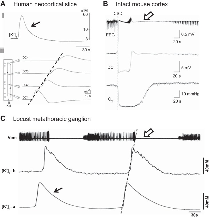
Fig. 1.
A locust model for cortical spreading depression. Ai: changes in extracellular K+ during a spreading depression-like event recorded in a human neocortical slice. Aii: propagating spreading depolarization (SD)-like events evoked by injection of 3 M KCl. Direct current (DC) potential measured at 4 locations in the slice (DC1–DC4). Velocity of propagation was 3.1 mm/min. B: combined measurement of EEG, DC potential, and Po2 during cortical spreading depression (CSD) in intact mouse cortex induced by pressure injection of 1 M KCl at the time of the dotted line. C: spreading depression-like events recorded in the metathoracic ganglion of a locust and induced by injection of 150 mM KCl. Vent, electromyographic recording of ventilatory motor pattern. Velocity of propagation was 1.9 mm/min. Note similarities in extracellular potassium ion concentration ([K+]o) surge (closed arrows), propagation of the event (broken lines), and period of electrical depression (open arrows) in the mammalian and insect preparations. A: adapted from Gorji et al. (2001); B: adapted from Takano et al. (2007); C: from Rodgers et al. (2007), CCBY 4.0, http://journals.plos.org/plosone/article?id=10.1371/journal.pone.0001366.

