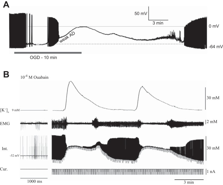
Fig. 3.
Neuronal responses to SD in rat midbrain and locust ganglion. A: whole cell recording of a midbrain neuron (locus ceruleus) of a rat in response to 10 min of oxygen/glucose deprivation (OGD), which induces a weak anoxic depolarization (weak AD). B: intracellular recording of a metathoracic ventilatory interneuron and [K+]o surges during repetitive SD induced with bath application of 1 mM ouabain. EMG indicates activity of a ventilatory muscle, and the current trace (Cur) indicates repetitive pulses for measurement of neuronal input resistance. Note that the mammalian neuron depolarizes by ∼60 to 0 mV, whereas for 19 insect neurons the membrane potential depolarized by only ∼15 to −35 mV. A: adapted from Brisson et al. (2014), CCBY 4.0, http://journals.plos.org/plosone/article?id=10.1371/journal.pone.0096585; B: adapted from Armstrong et al. (2009).

