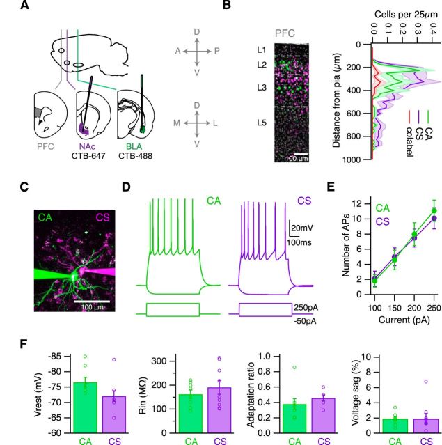
Figure 1.
Properties of CA and CS neurons in the infralimbic PFC. A, Schematic of injecting CTB-Alexa-647 into the NAc and CTB-Alexa-488 into the BLA of wild-type mice, to label neurons in the infralimbic PFC. Cardinal axes pictured to right for injection schematics apply to all figures. B, Left, CA (green) and CS (purple) neurons in the PFC, with DAPI staining in gray. White lines indicate laminar borders. Right, Quantification of CA, CS, and colabeled (red) cells in the PFC. C, Two-photon image of neighboring CA and CS neurons. D, AP firing and hyperpolarization in response to 250 pA and −50 pA current injections in the presence of synaptic blockers. E, Summary of AP firing over a range of current injections. F, Summary of resting membrane potential, input resistance, adaptation ratio, and voltage sag.

