
Figure 5.
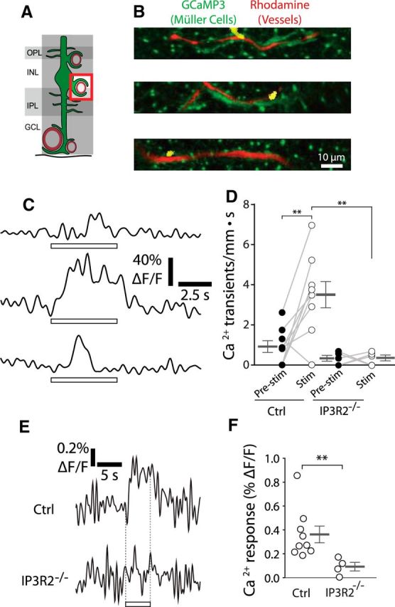
Stimulus-evoked Ca2+ signaling occurs in Müller cell endfeet contacting intermediate layer capillaries. A, Calcium signaling was monitored in Müller cell endfeet contacting intermediate layer capillaries. GCL, Ganglion cell layer; INL, inner nuclear layer; IPL, inner plexiform layer; OPL, outer plexiform layer. B, Examples of endfoot Ca2+ transients along intermediate layer capillaries. Imaging is the same as in Figure 3B. Similar images were obtained for 45 Ca2+ transients from nine eyecups. C, Traces of the Ca2+ transients in B. D, The frequency of endfoot Ca2+ transients (transients per millimeter of capillary length per s) increased during stimulation in control mice (Ctrl, p = 0.0046). Stimulus-evoked (stim; p = 0.0044) transient frequency was reduced in IP3R2−/− mice (n = 9 control eyecups, 5 IP3R2−/− eyecups; two-way ANOVA with Bonferroni posttest; control pre-stim, 0.91 ± 0.29 Ca2+ transients/mm · s; control stim, 3.5 ± 0.65 Ca2+ transients/mm · s; IP3R2−/− pre-stim, 0.32 ± 0.065 Ca2+ transients/mm · s; IP3R2−/− stim, 0.34 ± 0.069 Ca2+ transients/mm · s). E, Mean stimulus-evoked Ca2+ activity across all endfeet in control and IP3R2−/− mice. F, Amplitude of stimulus-evoked Ca2+ responses in E (n = 9 control eyecups, 4 IP3R2−/− eyecups; Mann–Whitney U test, p = 0.0028; control, 0.36 ± 0.070% ΔF/F; IP3R2−/−, 0.092 ± 0.035% ΔF/F). **p < 0.01.