
Figure 7.
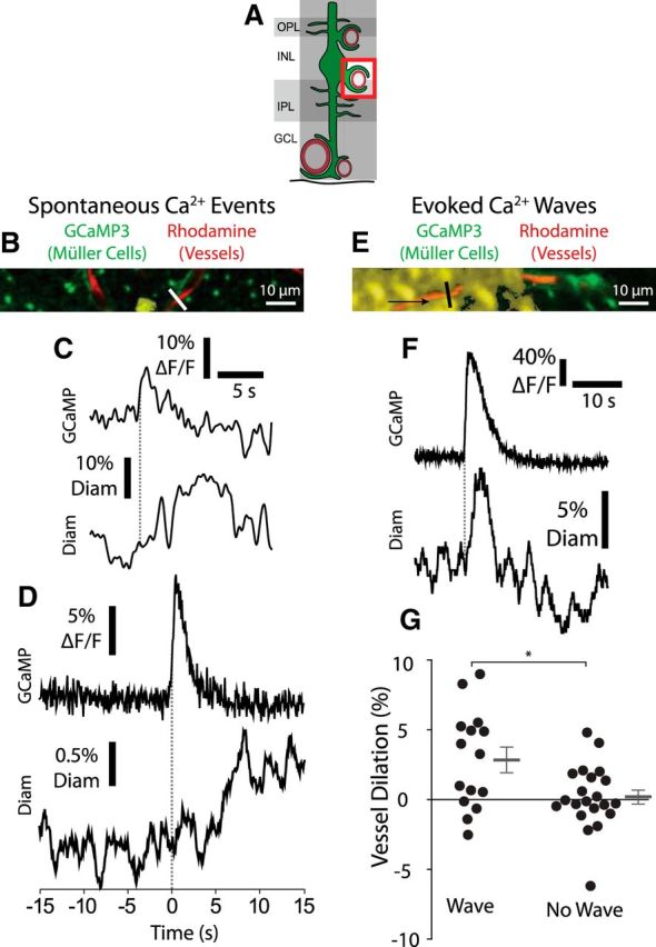
Calcium signaling in Müller cell endfeet is sufficient to evoke capillary dilation. A, Müller cell Ca2+ signaling and capillary diameter were monitored in the intermediate capillary layer. GCL, Ganglion cell layer; INL, inner nuclear layer; IPL, inner plexiform layer; OPL, outer plexiform layer. B, Example of a spontaneous endfoot Ca2+ transient along an intermediate layer capillary. Labeling is the same as in Figure 3B. Similar images were obtained for 207 Ca2+ transients from five eyecups. C, Trace of the Ca2+ transient in B (top), along with the resulting change in capillary diameter (bottom; measured at the white line in B). The onset of the Ca2+ transient is indicated by the dotted line. D, Mean intensity of spontaneous Ca2+ transients along with the mean change in capillary diameter. Capillary diameter increased following Ca2+ transients (n = 208 transients from 5 eyecups; Wilcoxon matched pairs signed rank test, p < 0.001; pre-transient, −0.1 ± 0.8% change in diameter; post-transient, 0.8 ± 0.8% change in diameter). Traces were synchronized to the rising phase of the Ca2+ transients (dotted line). E, Example of a Müller cell Ca2+ wave (yellow), generated by a brief negative current pulse from a glass pipette, propagated in the direction indicated by the arrow. Similar images were obtained for 14 Ca2+ waves from four eyecups. F, Endfoot Ca2+ signaling (top) and capillary diameter (bottom) as the Ca2+ wave in E propagated past the capillary. Endfoot Ca2+ and vessel diameter were measured at the black line in E. G, Mean change in capillary diameter when a Ca2+ wave reached a capillary and when the Ca2+ wave failed to reach the capillary (n = 15 Ca2+ waves and n = 21 trials without Ca2+ waves from 4 eyecups; unpaired t test, p = 0.0095; with Ca2+ wave, 2.8 ± 0.90% vessel dilation; without Ca2+ wave, 0.19 ± 0.50% vessel dilation). *p < 0.05.