Figure 1.

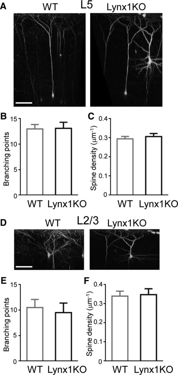
Normal dendritic complexity and spine density of L5 and L2/3 V1 neurons in adult Lynx1-KO mice. A, 3D reconstruction of in vivo two-photon images of L5 pyramidal neuron in binocular zone of V1 of adult WT and Lynx1-KO mouse. Scale bar, 100 μm. B, Number of branching points of apical dendrite of L5 neurons (WT: n = 7 cells from 5 mice, KO: n = 9 cells from 6 mice). C, Spine density (number of spines per micrometer) of L5 neurons (WT: n = 11 cells from 5 mice, KO: n = 13 cells from 8 mice). D, 3D reconstruction of two-photon images of L2/3 pyramidal neurons. E, Number of branching points of apical dendrite of L2/3 neurons (WT: n = 6 cells from 4 mice, KO: n = 4 cells from 3 mice). F, Spine density (number of spines per micrometer) of L2/3 neurons (WT: n = 10 cells from 5 mice, KO: n = 10 cells from 7 mice). One to three dendrites were imaged from each cell to count the total spines in each cell. Data are presented as mean ± SEM.
