Figure 2.

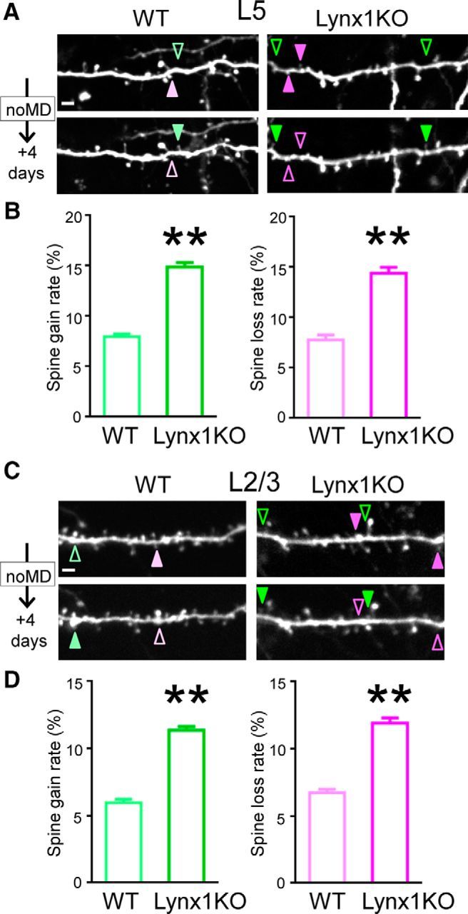
Spine turnover of adult L5 and L2/3 V1 neurons is higher in Lynx1-KO mice than in WT mice. A, Repeated imaging of dendritic segments of adult L5 V1 pyramidal neurons over 4 d in adult WT mice and Lynx1-KO mice. Light green (WT) and green (KO) arrowheads indicate gained spine. Light magenta (WT) and magenta (KO) arrowheads indicate lost spine. Scale bar, 5 μm. One to three dendrites were imaged from each cell to count total, gained, and lost spines in each cell. B, Spine gain and loss rate of adult L5 V1 pyramidal neurons over 4 d are significantly higher in adult Lynx1-KO mice than in WT mice (WT: n = 11 cells from 5 mice, KO: n = 13 cells from 8 mice). C, Repeated imaging of dendritic segment of adult L2/3 V1 pyramidal neuron. D, Spine gain and loss rate of adult L2/3 V1 pyramidal neurons over 4 d were significantly higher in adult Lynx1-KO mice than in WT mice (WT: n = 10 cells from 5 mice, KO: n = 10 cells from 7 mice). Data are presented as mean ± SEM. **p < 0.01.
