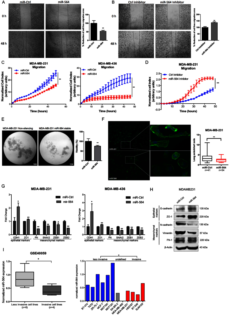
Figure 2. miR-564 inhibits migration and invasion of breast cancer cells by blocking EMT.
(A) In vitro scratch assay with MDA-MB-231 cell line at 0 and 48 hours post-transfection with miR-564 or control miRNA. (B) Wound healing assay in MDA-MB-231 cell line at 0 and 48 hours post-transfection with miR-564 inhibitor or control hairpin inhibitor. Quantification was performed by the measurement of gap distance after cell migration. (C) Real-time migration assay using RTCA with MDA-MB-231 and MDA-MB-436 cells in the presence of miR-564 or control miRNA. (D) Real-time migration assay using RTCA with MDA-MB-231 cells upon miR-564 antagonist. (E) Poly-HEMA assay to analyze anchorage-independent growth of MDA-MB-231-luc cells stably-transfected with miR-564 and incubated for 48 hours. Control group was represented by cells stably-transfected with empty vector. Tumorigenicity was quantified as percent viable cells measured with WST-1 cell proliferation reagent. (F) DAPI/Phalloidin staining of MDA-MB-231 cell line. Images were taken 24 hours after transient transfection with miR-564 mimic. Cell morphology changes were quantified as ratio of long axis to short axis for each cell. (G) qRT-PCR showing the expression of epithelial and mesenchymal markers in MDA-MB-231 and MDA-MB-436 cell lines upon miR-564 mimic transfection. (H) Western blot showing changes in protein levels of EMT markers in miR-564 transfected MDA-MB-231 cells. (I) Expression of miR-564 in invasive and less-invasive breast cancer cell lines from GSE40059 dataset (left panel). Expression in each cell line is depicted as relative expression with respect to MDA-MB-468 (right panel).

