FIG 3 .

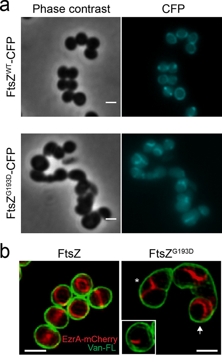
Fluorescent fusions with FtsZ and EzrA lose mid-cell localization in M5 cells. (a) Localization of FtsZWT-CFP in BCBAJ020 cells (top) and FtsZG193D-CFP in BCBRP005 cells (bottom). Phase-contrast (left) and epifluorescence (right) images are shown. (b) SIM images of EzrA-mCherry (red) and cell wall labeled with Van-FL (green) in BCBAJ012 cells expressing FtsZWT (left) and in BCBRP006 cells expressing FtsZG193D (right). In the presence of FtsZG193D, EzrA-mCherry fails to localize as a mid-cell ring. Instead, structures assemble on only one side of the cell (white box) or in Y patterns compatible with one-turn helical structures (asterisk). More elongated BCBRP006 cells show EzrA-mCherry localized toward the poles of the cell (white arrow). Scale bars: 1 µm.
