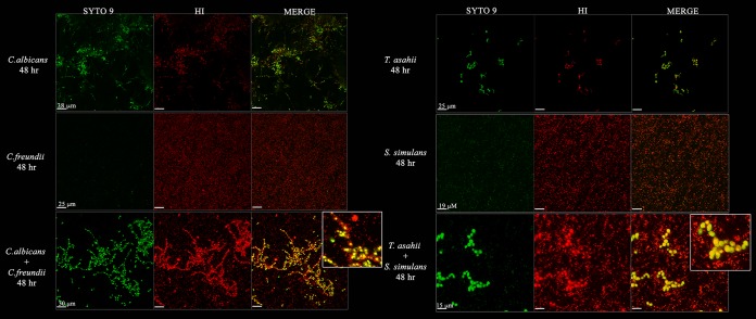
FIG 5 .
Pathogens form interkingdom biofilms. Fluorescent confocal microscope images of mono- or coculture Candida albicans and Citrobacter freundii or Trichosporon asahii and Staphylococcus simulans. Biofilms were grown for 48 h at 37°C on polystyrene plates in RPMI 1640 medium with GlutaMAX supplement, washed to remove planktonic cells, and stained with SYTO 9 and hexidium iodide (HI) prior to imaging. Fungi (large) and bacteria (small) can be distinguished by size. Fungi appear green and bacteria appear red in the merged images. The size of the scale bar for each row is labeled in the first column (SYTO 9) for each culture. The insets are zoomed-in portions of the merged coculture biofilm images.

