
Fig. 5.
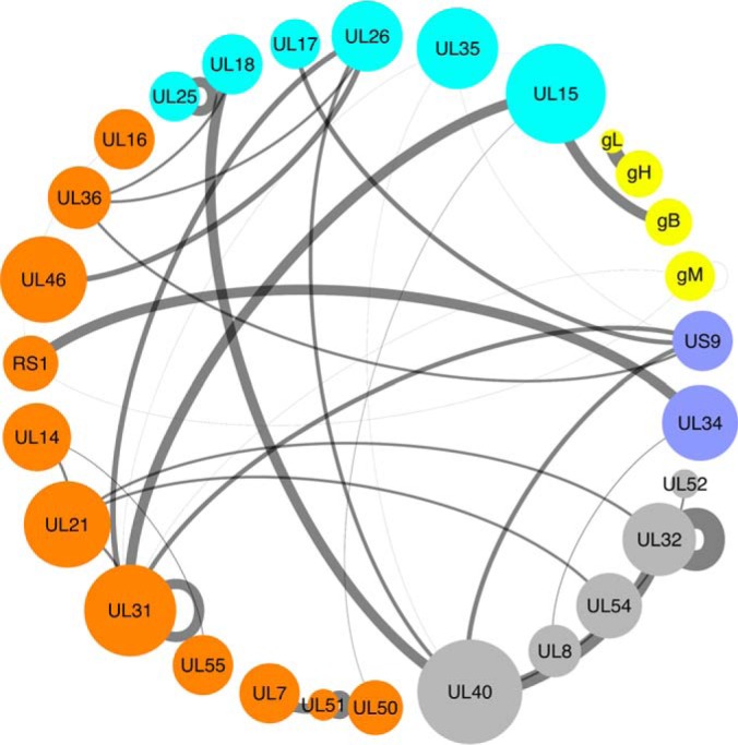
Subnetwork highlighting homologous PPIs with a MIscore > 0. 4 (supplemental Table S2). The node size indicates the number of interacting partners for each protein (degree). Edge width is scaled according to the confidence score associated with the PPI. The nodes are color-coded according to the protein location in the virion particles - gray: protein has not been detected in virions by MS in Loret et al. (34); cyan - capsid and capsid-associated protein; orange - tegument protein; yellow - envelope glycoprotein; dark blue - envelope protein (not glycoprotein).