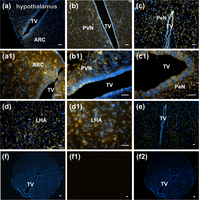
Figure 1.
Localization of nesfatin-1-immunopositive cells in hypothalamic nuclei (a–e) in male rats. (a) and (a1), arcuate nucleus (ARC); (b) and (b1), paraventricular nucleus (PVN); (c) and (c1), periventricular nucleus (PeN); (d) and (d1), lateral hypothalamic area (LHA); (e), whole hypothalamic area; (f), (f1,f2), negative control. TV: third ventricle, bar = 100 μm.

