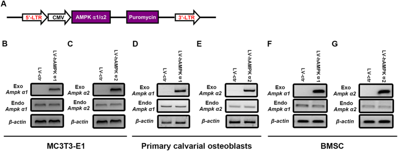
Figure 2. Generation of MC3T3-E1, primary osteoblasts and mouse BMSCs stably over-expressing AMPK α1 and α2 subunits.
The AMPK α1 and α2 subunit coding sequences were cloned into an LV vector with a CMV promoter and positive clones were selected with puromycin (A). An empty LV vector served as a control (LV-ctr). RT-PCR was employed to check the expression of exogenous and endogenous α1 (B,D,F) and α2 (C,E,G) subunits of AMPK was evaluated. β-actin was used as an internal control.

