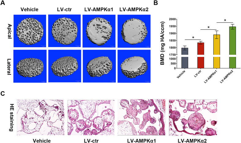
Figure 4. In vivo ectopic bone formation in MC3T3-E1 cells over-expressing AMPK α1 and α2.
β-TCP scaffolds loading with and without MC3T3-E1 cells over-expressing AMPK α1 and α2 were implanted into the intramuscular pocket of the femur of nude mice. Eight weeks later, the complexes were harvested and scanned by micro-CT (A). Micro-CT images are shown in apical and lateral views. The measurement of BMD was performed on the basis of micro-CT (B). Then these complexes were decalcified, embedded in paraffin and stained with hematoxylin and eosin (H&E). Data were shown as the mean ± SD. *p < 0.05.

