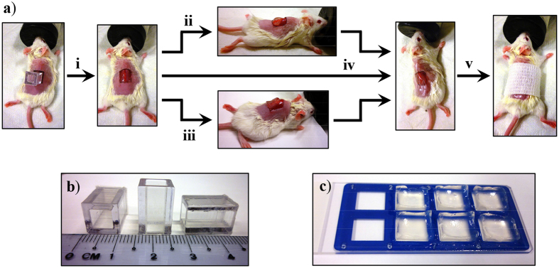
Figure 4.
(a) Schematic of the full-thickness excision mouse model used to evaluate the wound healing property of LK6C + CRGD hydrogel dressings. (i) The wound boundaries were marked out on dorsally shaved mice using a template (as shown in b). Both the epidermis and dermis were then surgically removed to create 1 × 1 cm full-thickness wounds. Either the (ii) positive control (DuoDerm hydroactive gel) or (iii) peptide hydrogel (cast as ready-to-use sterile gel blocks, as shown in c) was applied to the wounds. (iv) All wounds were then covered up with Tegaderm, including the negative control group which received no gel application. (v) The wound was lightly bandaged and left to heal. Animals were sacrificed on day 3, 7 and 14 for histological examination.

