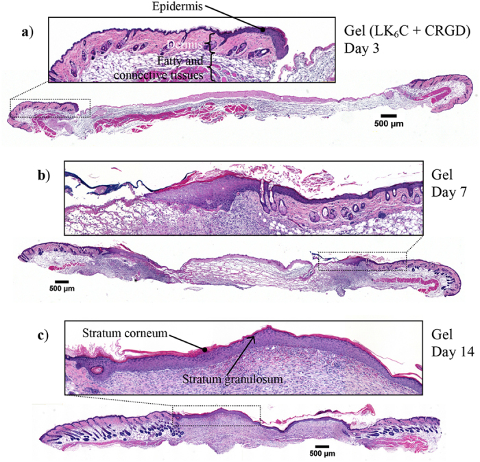
Figure 5. Representative cross-sectional histological sections of wounds treated with peptide hydrogel infused with completed medium.

Skin samples were obtained on day (a) 3, (b) 7, (c) 14 for staining with H&E. Over time, the leading front of the epidermal “tongue” was observed to migrate inwards to close up the wound and restore the critical barrier function of skin. Re-epithelialization was completed by day 14, along with the appearance of a visible layer of stratum corneum and stratum granulosum. All insets show magnified images of the respective boxed regions.
