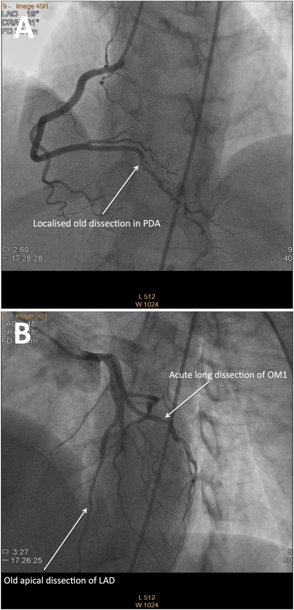
Figure 3.

Multiple recurrent spontaneous coronary artery dissections of varying chronicity in one patient. (A) Coronary angiography of the posterior descending artery (PDA) demonstrates diffuse irregularity with a focal ulceration or dissection in its first centimetre from original presentation 2 months prior. (B) Coronary angiography of the left anterior descending artery demonstrates an old healing dissection of the terminal segment and a new dissection of a single large obtuse marginal (OM) branch of the circumflex.
