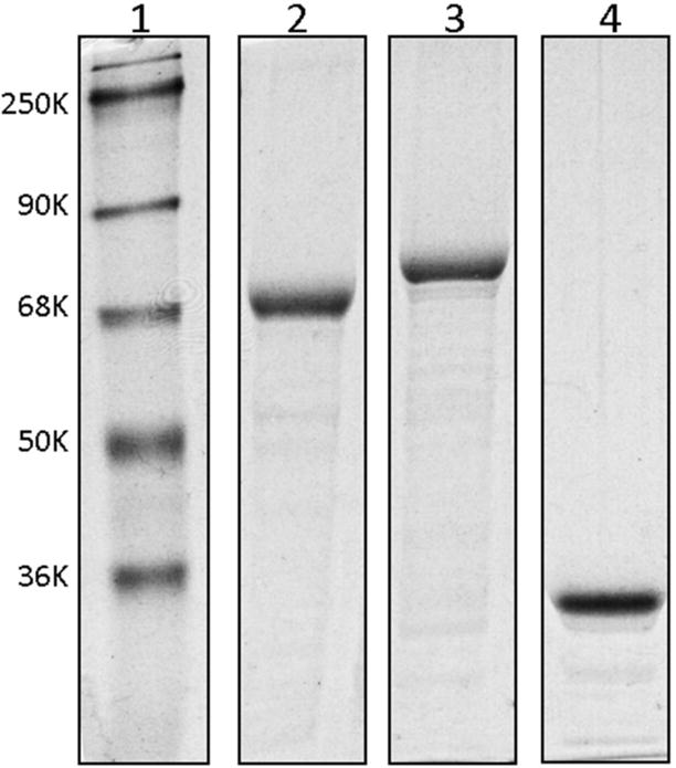
Fig. 3. SDS-PAGE analysis of various cdb3-eGFP constructs following expression and purification from bacteria.

Cdb3-eGFP proteins were expressed in E. coli strain BL21(DE3) pLys S, purified using an affinity column, and analyzed by SDS-PAGE: lane 1, molecular markers; lane 2, human cdb3-eGFP fusion protein containing human cdb3 residues 1–379; lane 3, mouse cdb3-eGFP fusion protein containing mouse cdb3 residues 1–398; lane 4, eGFP protein which is not fused with cdb3.
