
Fig. 5. Measurement of Hb binding to cdb3-eGFP by analysis of FRET.
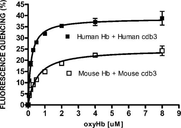
Increasing concentrations of Hb (0.07μM to 8μM) were incubated for 30 min at room temperature with cdb3-eGFP (0.2μM) in 10 mM BisTris buffer (pH6.5). Emission spectra were then recorded and the fluorescence intensity at 510nm was used to calculate the % fluorescence quenching using the equation described in the text. Prism software was employed to determine a Kd of human oxyhemoglobin for human cdb3 of 1.8 × 10⁻⁷ M; and the Kd of murine oxyhemoglobin for murine cdb3 of 5.5 × 10⁻⁷M. The data constitute the mean +/− S.D. of two experiments.