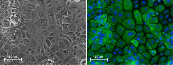
Fig. 1.

Isolation and culture of primary HBEC. HBEC are shown in a transmitted light image (left panel) and immune-stained for pan-cytokeratin (right panel, green = cytokeratin, blue = nuclear DNA). The antibodies used were anti-human pan-cytokeratin rabbit polyclonal (Life Technologies, DP010-05) and anti-rabbit IgG –Cy3 conjugate (Jackson Immuno Research Laboratories, 711-166-152). Bar = 100 μm
