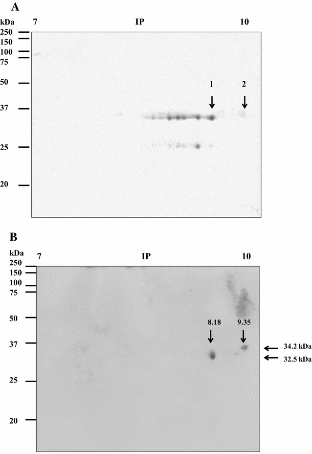
Figure 3.

Separation of the M. haemolytica outer membrane proteins that bind to bovine apolactoferrin (BapoLf) using 2-D gel electrophoresis. A 2-D gel electrophoresis of the M. haemolytica OMP, stained with Coomassie blue. B Overlay of M. haemolytica OMP, from 2-D gel electrophoresis; the nitrocellulose membrane was incubated with HRP-BapoLf. The arrows show the spots of BapoLf binding proteins (A) and their MW and estimated IP (B), which were further identified by mass spectrometry.
